Eye Care Technology
California
Developed for medical imaging and is a standard for retinal screening programs. California is available in multiple configurations and multiple imaging modality options. Californiaproduces a 200°, single-capture retinal image of unrivaled clarity in less than ½ second and is changing the management of diseases including Geographic Atrophy, DR, AMD, and uveitis.
With California, Optos has incorporated new technology enabling practitioners to see more, discover more, and effectively treat more ocular pathology thus promoting patient health. In addition to a field of view of 200 degrees or 82% of retina in a single image capture, California, offers the following benefits:
Visibility of 50% more of the retina when compared to other conventional imaging devices
Motorized head and chin rest to more easily align those patients who require additional assistance during imaging
Multiple imaging modalities including color rg which produces three images in a single capture (color rg, sensory red-free, and choroidal in a single image) and introducing color rgb which produces four images in a single capture (color rg, color rgb, sensory red-free, and choroidal) and autofluorescence (both green and blue*) (*Feature may not be available in all regions)
Dye-based imaging modalities fa and icg as well as interweaved angiography for parallel capture of fa and icg images without manually switching between imaging modalities
For more information please check out: optos.com

Zeiss Cirrus 6000
CIRRUS® 6000 is the next-generation OCT from ZEISS, delivering high-speed image capture with HD imaging detail and a wider field of view so you can make more informed decisions and spend more time with the patients who need it.
Faster, wider with a new level of detail
At 100,000 scans per second, ZEISS CIRRUS 6000 enables clinicians to image a larger field of view up to 12mm in a single scan. It also captures high-definition (HD) OCT and OCT Angiography (OCTA) scans, revealing the finer microvascular details of the retina and providing more insight into your patient’s condition.
For more information please check out: Zeiss.com

Zeiss Perimetry
There's a Humphrey Field Analyzer compatible with every practice ready to deliver the highest standard of care in the detection and management of glaucoma.
For more information please check out: zeiss.com

Humphrey Matrix 800
Operating a visual field instrument doesn’t get much easier than a Humphrey Matrix. It provides the ideal solution for busy practices for case detection and fast threshold testing. In addition to simplifying visual field testing, numerous studies show that frequency doubling perimetry can detect visual field loss missed by other methods.
For more information please check out: Zeiss.com

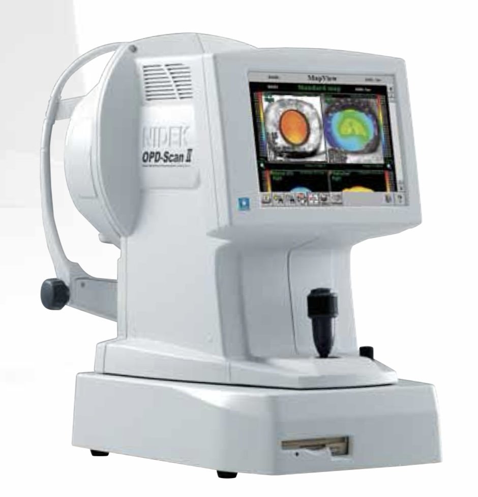
OPD-Scan II ARK-10000
OPTICAL PATH DIFFERENCE SCANNING SYSTEM
The NIDEK OPD-Scan II provides information on corneal topography, wavefront, autorefraction, keratometry and pupillometry in one unit, utilizing state-of-the-art imaging and analysis technology developed specifically to measure normal to highly aberrated eyes. The system offers a variety of data maps to provide information on the total refractive error, wavefront, corneal shape, internal aberrations and visual quality of the eye, allowing highly accurate and reliable information for optic diagnostics.
For more information please check out: medlikim.com